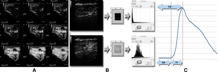
Fig. 1.
Example of a CEUS examination of a soft-tissue mass. a Patient with soft-tissue metastases: representative images from various contrast-enhancement phase. b Selection of an ROI (0.25 cm × 0.25 cm) in CEUS image that is representative of the lesion. c Contrast-enhancement parameters extrapolated from the time–enhancement intensity curve

