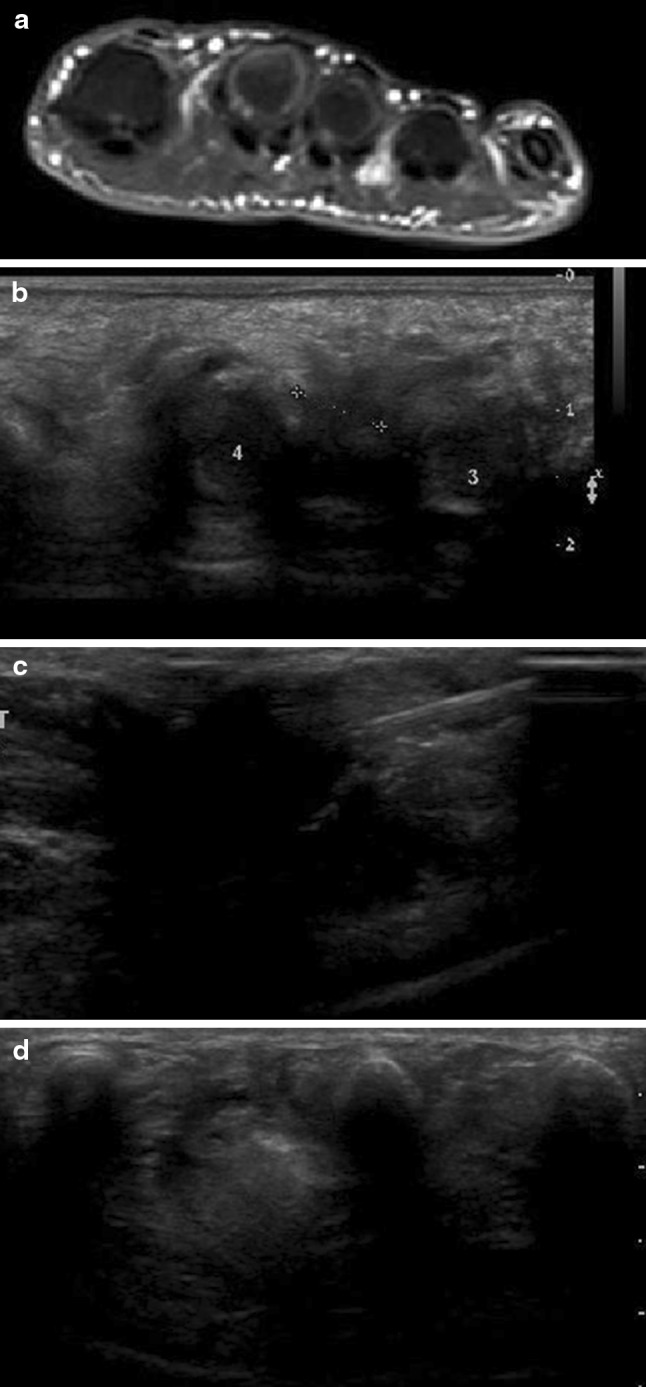
Fig. 2.

Morton’s neuroma. Axial STIR T2-weighted MRI scan. The neuroma is clearly visualized in the space between the third and fourth metatarsals (a). Scan performed along the short axis of the nerve with a high-frequency linear transducer. The neuroma is clearly visualized as a hypoechoic lesion located in the space between the metatarsal heads (b). The needle tip is inserted into the neuroma (c). After treatment, the neuroma appears hyperechoic (d)
