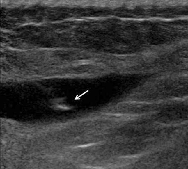
Fig. 4.

Intralesional flow. US reveals an intramuscular hematoma whose content appears hypoechoic. Injection of normal saline is associated with a hyperechoic ‘spurt’ of fluid within the lesion (arrow), which confirms that the needle tip has been correctly placed
