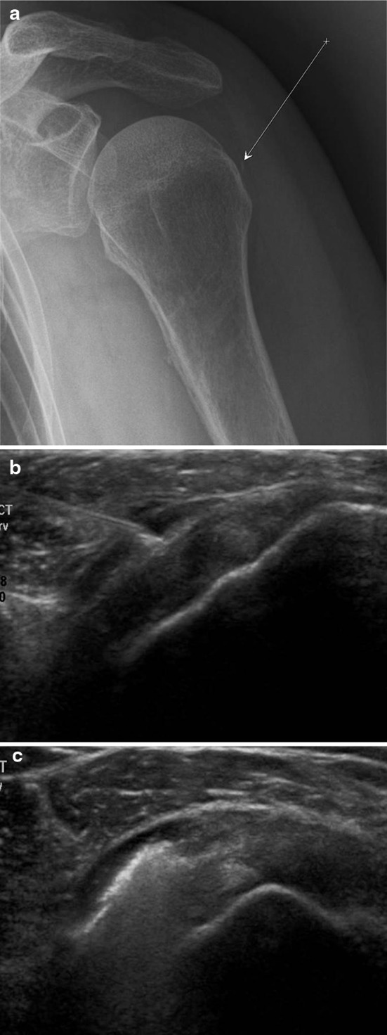
Fig. 7.

US-guided infiltration of the subdeltoid bursa. Anteroposterior radiograph: the arrow identifies the trajectory of the needle and the arrowhead the bursa (a). Coronal US scan performed with a high-frequency linear array transducer. The needle is advanced into the bursa, which is located between the deltoid muscle and the rotator cuff (b). Coronal US scan performed with a high-frequency linear array transducer. The image confirms uniform distribution of the injected substance within the bursa (c)
