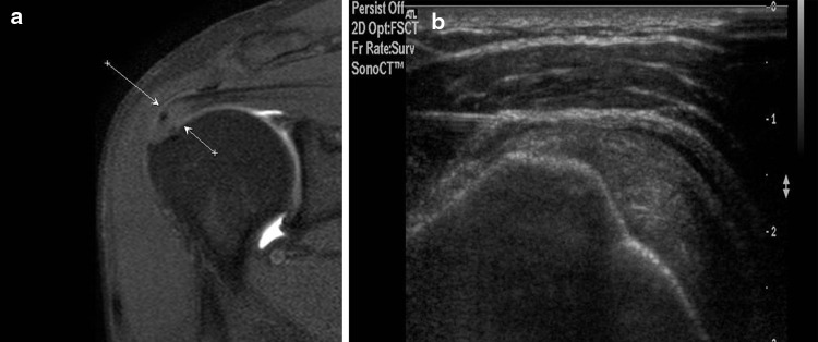
Fig. 8.

US-guided infiltration of enthesitis of the supraspinatus tendon. Coronal MRI scan (proton density weighted with fat suppression): the arrows indicate an area of degenerative changes at the insertion of the supraspinatus tendon (a). US scan along the long axis of the tendon with a high-frequency linear array transducer (b). The tip of the needle is visualized as it is advanced toward the degenerated zone of the tendon
