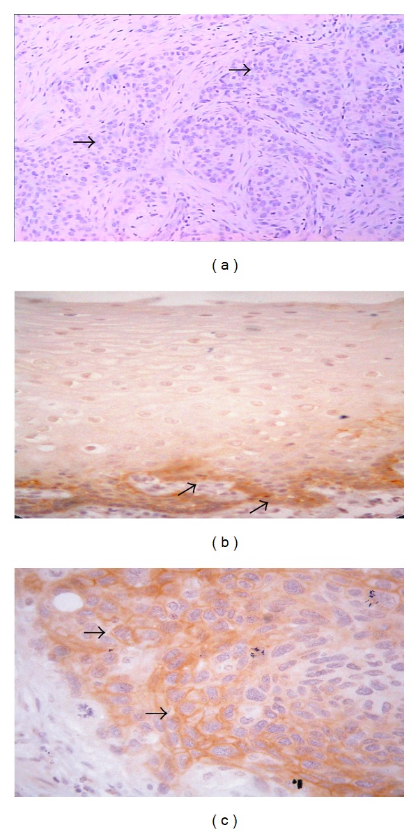
Figure 1.

A negative control (a breast cancer known for positive FXYD-3), where the primary FXYD-3 was replaced by PBS, showed no staining of the FXYD-3 in tumor cells (→) (a). Immunohistochemical assay for FXYD-3 expression in esophageal squamous cell carcinoma (ESCC). FXYD-3 was weak expression in normal mucosa, mainly located in the basal cell layer (→) (b), and strong expression in primary tumor cells (→) (c).
