
Figure 1.
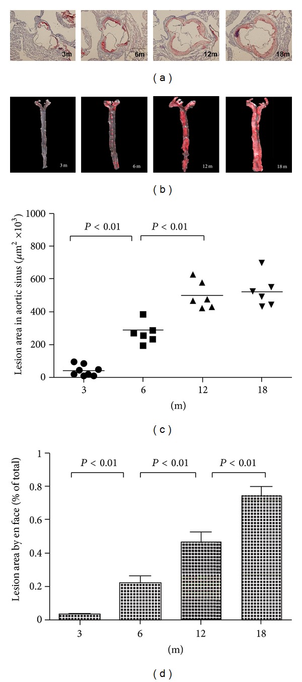
Atherosclerotic lesion areas in proximal aortas of apoE−/− mice with different ages. (a) Representative atherosclerotic lesions in cross-sections of proximal aortas stained with oil-red O. (b) Representative en face aortas from 3 months to 18 months. Quantitation of the mean lesion area of aortic sinus (c) and percentage lesion area in the aorta by en face analysis (d).