
Figure 2.
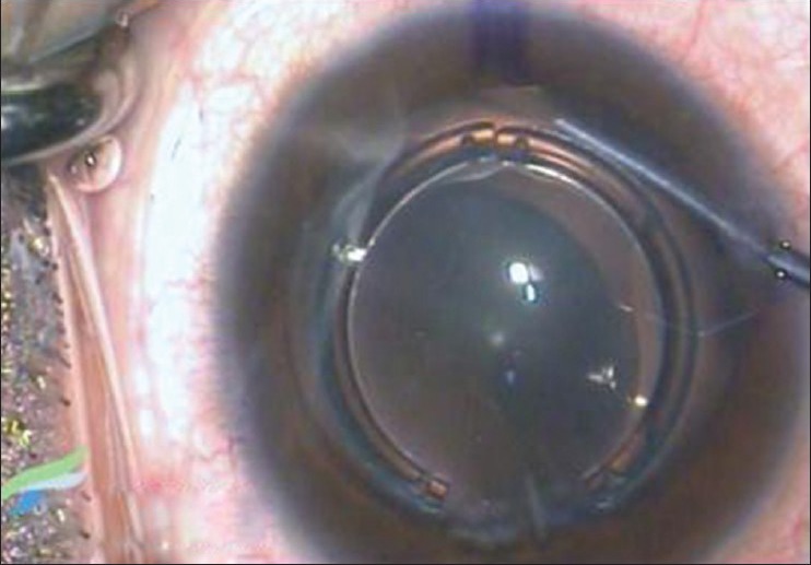
Intraoperative snapshot of the TICL haptic being tucked into the ciliary sulcus. The intrastromal corneal rings can be seen in situ in the cornea
Official websites use .gov
A
.gov website belongs to an official
government organization in the United States.
Secure .gov websites use HTTPS
A lock (
) or https:// means you've safely
connected to the .gov website. Share sensitive
information only on official, secure websites.
Intraoperative snapshot of the TICL haptic being tucked into the ciliary sulcus. The intrastromal corneal rings can be seen in situ in the cornea