Figure 1.
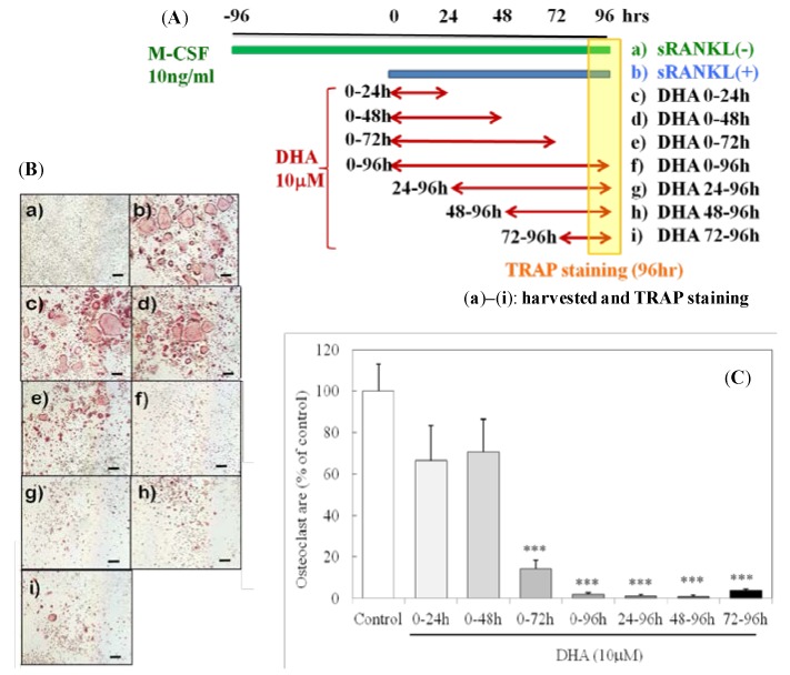
DHA inhibited sRANKL-induced osteoclast differentiation at the late stage. (A) Culture conditions and (B) representative images of osteoclasts (TRAP staining (a)–(i)). The scale bar indicates 200 μm. (C) The areas occupied by osteoclasts (TRAP+ cells with three or more nuclei) were analyzed. Each column and bar represents the mean ± SE of four to five wells. *** Significantly different from the control (sRANKL(+)); p < 0.0001 by Dunnett’s multiple comparison test.

