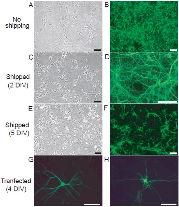
Figure 2. Neuronal viability decreases with increased age of primary hippocampal cultures at the time of shipping.
Phase contrast (A, C, E) and fluorescent (B, D, F, G and H) microphotographs of primary cultures of dissociated hippocampal neurons at 7DIV. All cultures were set up from the same dissection of postnatal day 1 (P1) rat hippocampi. A subset of cultures was transfected with MAP2-eGFP at 4 DIV (G, H); remaining cultures were immunostained for MAP2 at 7 DIV (B, D, F). Relative to control cultures that were not shipped (A, B), shipping cultures at 2 DIV (C, D) had minimal impact on neuronal cell viability whereas shipping cultures at 5 DIV (E, F) significantly decreased neuronal cell health. Comparison of transfected neurons in cultures that were not shipped (G) versus those in cultures shipped at 5 DIV confirms decreased viability in the latter. Bar = 50μm.

