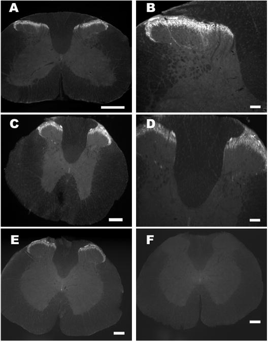
Fig. 2.

Fluorescence images of rat spinal segments labeled with phoenixin antiserum or phoenixin antiserum pre-absorbed with the peptide. A, C and E, cervical, thoracic and thoracolumbar spinal segment where irPNX is detected in superficial layers of the dorsal horn. B and D, a high magnification of A and C, where several strands of irPNX cell processes extend into the deeper laminae. F, a thoracolumbar segment labeled with phoenixin-antiserum pre-absorbed with the peptide (1 μg/ml); irPNX is not detected in this spinal section. Scale bar: A, C E and F, 250 μm; B and D, 100 μm.
