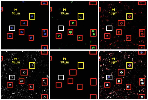
Fig. 4.
Representative, confocal images from a negatively depleted blood sample from a patient with stage IV triple negative breast cancer. Markers shown are with DAPI/nuclei, FITC/cytokeratins, AF594/vimentin, and APC/CD44. In the composite, the cells in red boxes are positive for all four markers, while cells encircled with yellow boxes are negative for cytokeratins, yet positive for the other three markers. White boxes are cells negative for cytokeratins and vimentin, and CD44, positive for DAPI/nuclei

