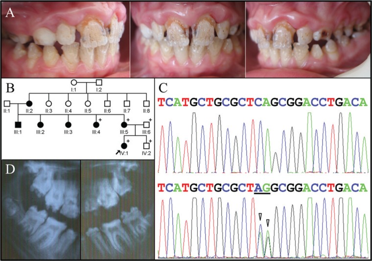
Figure 2.
Clinical and mutational analysis of Family 2. (A) Frontal and lateral clinical photographs of the proband, showing maxillary anterior gingival hypertrophy and generalized severe enamel hypoplasia with deep pits and grooves, often oriented vertically. (B) Pedigree of Family 2. The “plus” symbol indicates members recruited for this study. (C) LAMB3 exon 23 sequencing chromatogram for the unaffected family member (IV:2) with the wild-type sequence (top) compared with the heterozygous mutated allele in the proband (IV:1) with a 2-bp substitution of CA with AG (underlined; c.C3431A, p.Ser1144*). (D) Radiographs of the posterior teeth showing severe enamel hypoplasia. The molar root form is within normal limits, with only a trace of taurodontism.

