
Fig. 3.
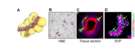
From the vascular niche to adipocyte. (A) Illustration conceptualizing the adipose vascular niche depicting blood vessels (red), adipose stem cells (green with green nuclei), mural cells (green with black nuclei) and mature adipocytes (yellow with green nuclei). (B) Mouse subcutaneous adipose tissue stained with Hematoxylin and Eosin (H&E) stain. (C) Immunofluorescence image of mouse subcutaneous adipose tissue section showing the expression of GFP (marking adipose stem cells, green), PECAM (an endothelial marker, red) and smooth muscle actin (SMA) (a mural cell marker, blue). (D) Stromal vascular particulates (SVPs) were isolated from mouse subcutaneous fat depots and immunostained for GFP (adipose stem cell, green), PECAM (endothelial marker, red) and SMA (mural cell marker, blue). Yellow arrows indicate adipose stem cells (green) residing at the vascular interface.