Fig. 4.
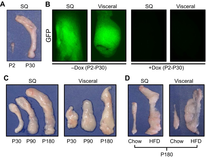
Fat expansion. (A) Isolated postnatal day (P)2 and P30 subcutaneous (inguinal) fat depots, demonstrating the extensive expansion that occurs within this short window of time. (B) Adipo-trak mice (Tang et al., 2008) were treated with placebo (-Dox) or doxycycline (+Dox) at denoted times. In the absence of doxycycline the GFP marker is expressed and the adipose depots are green. In the presence of doxycycline, all GFP signal is lost, indicating that there was a massive recruitment of new stem cells and adipocyte replacement. (C) White adipose depots enlarge with age; subcutaneous and visceral fat depots were isolated from 30-, 90- and 180-day-old mice. Depot weight nearly quintupled between 30 and 180 days. (D) Subcutaneous inguinal (SQ) and visceral perigonadal depots obtained from mice on a standard diet (chow) or a high fat diet (HFD), demonstrating the dramatic expansion in response to HFD.

