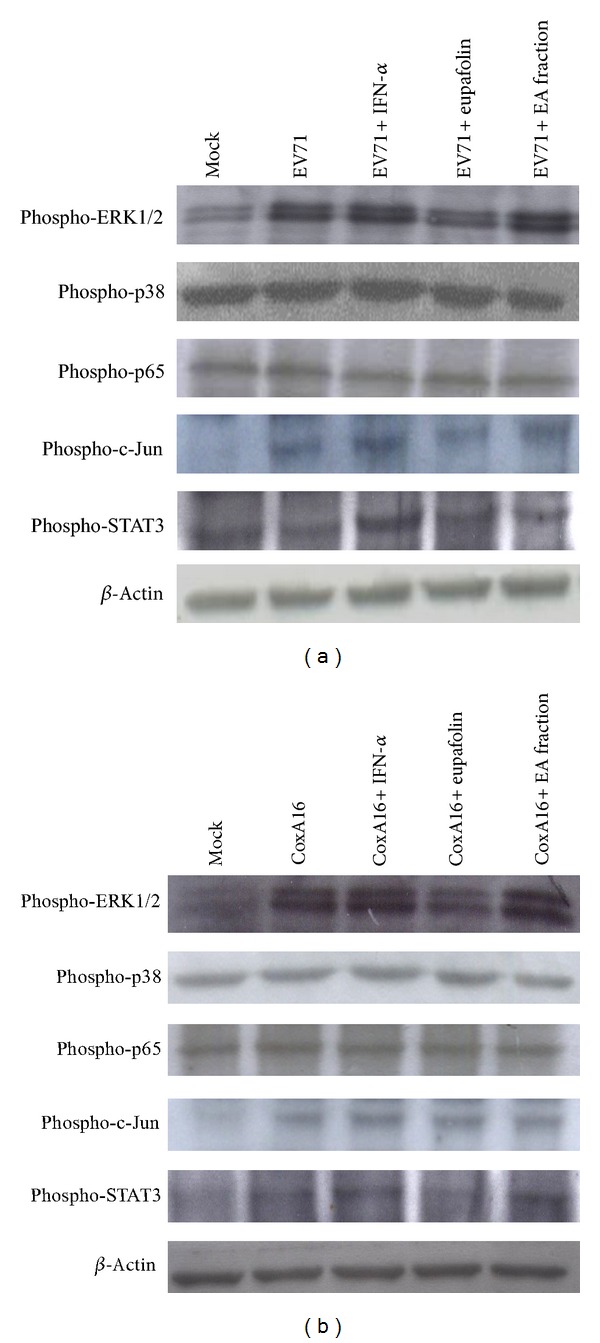
Figure 10.

Phosphorylation levels of ERK1/2, p38 MAPK, p65 (NF-κB), c-Jun, and STAT3 in infected RD cells treated with(out) eupafolin and EA fraction. EV71 (a) or CoxA16 (b) infected RD cells were harvested 9 h posttreatment, and lysates resolved on 10% SDS-PAGE and transferred onto nitrocellulose paper. Blot was probed with specific mAbs, developed with alkaline phosphatase-conjugated secondary antibody and enhanced chemiluminescence substrates. Lane 1, mock cells; Lane 2, infected cells; Lane 3, infected cells treated with IFN-α; Lane 4, infected cells treated with eupafolin; Lane 5, infected cells treated with EA fraction.
