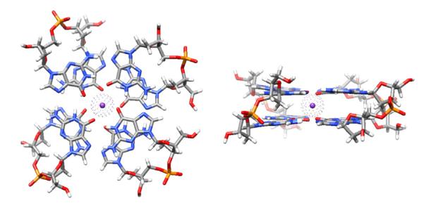
Fig. 1.

Left – 2-quartet G-DNA stem with anti-parallel down-down-up-up (from bottom left counterclockwise) 5′-GpG-3′ strand orientation and with anti-syn bases in 5′-3′ direction in all strands. The structure is colored by atom types with the integral bound K+ ion shown with a van der Waals sphere. Right – side view.
