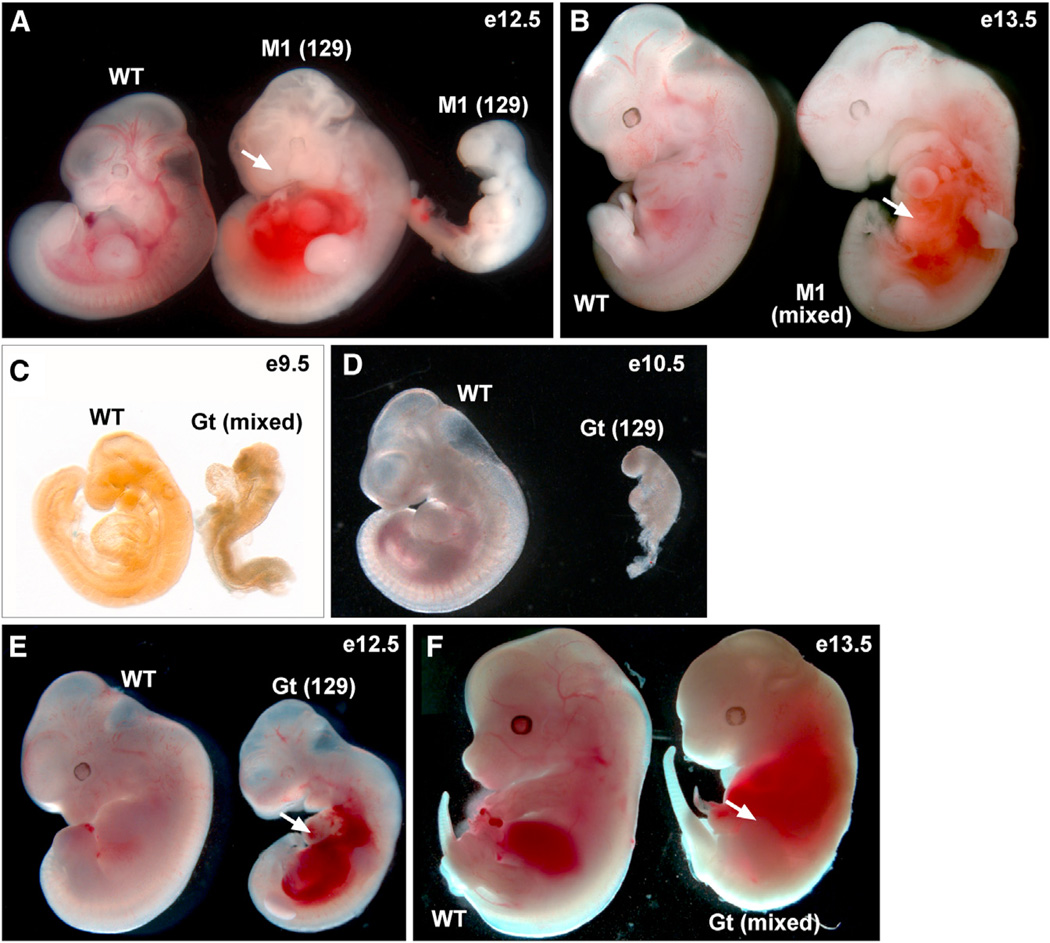
Figure 1. Similar Phenotypes of Senp1 Mutants.
(A) An E12.5 WT embryo and two Senp1M1/M1 (M1) littermates on 129S6 (129) inbred background. The mutant embryo in the middle shows pooling of blood in the abdominal region (arrow). Mutant embryo at right arrested at E11.5 or earlier.
(B) 129S6/C57Bl6 background (mixed) E13.5 WT embryo and M1 littermate, showing pooling of blood in the abdominal region (arrow).
(C) Mixed background E9.5 WT embryo and Senp1Gt/Gt (Gt) littermate. The mutant arrested prior to embryonic turning.
(D) 129S6 background E10.5 WT embryo and Gt littermate. The mutant arrested earlier.
(E) 129S6 background E12.5 WT embryo and Gt littermate. The mutant is developmentally delayed and shows pooling of blood in the abdominal region (arrow).
(F) Mixed background E13.5 WT and Gt littermate. The mutant shows pooling of blood in the abdominal region (arrow).
See also Table S1.

