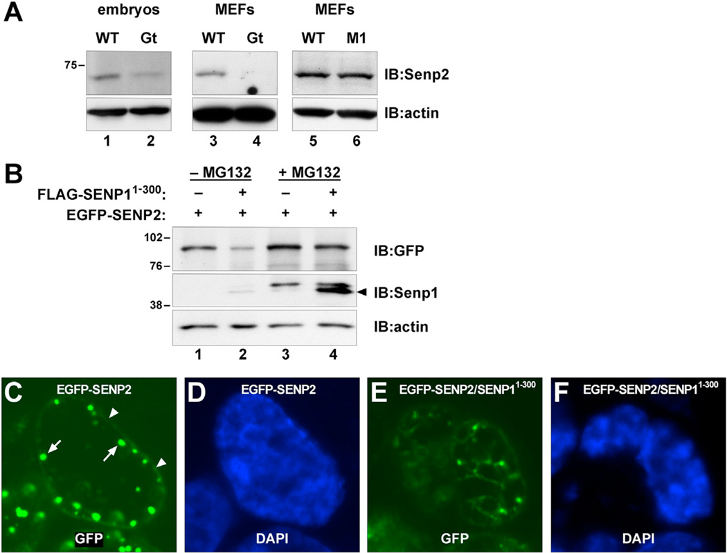
Figure 4. Truncated Senp1 Affects Senp2 Expression.
(A) Immunoblots of lysates from E10.5 Gt mutant embryos (lanes 1 and 2) or MEFs (lanes 3 and 4), or M1 mutant MEFs (lanes 5 and 6) detected with anti-Senp2. Senp2 levels are significantly lower in Gt but are not affected in M1.
(B) Immunoblots of HEK293 cells transfected with EGFP-SENP2 fusion alone (lanes 1 and 3) or co-transfected with FLAG-SENP11–300 (lanes 2 and 4), detected with anti-GFP (upper panel) or anti-Senp1 (middle panel). EGFP-SENP2 levels are significantly reduced in the presence of FLAG-SENP11–300 (lane 2). Reduced levels are no longer seen following MG132 treatment (lane 4), indicating that FLAG- SENP11–300 enhances proteasomal turnover of EGFP-SENP2. Lower panel shows anti-actin to control for equal loading.
(C) EGFP fluorescence showing that transfected EGFP-SENP2 localizes predominantly around the nuclear rim (arrowheads) and within subjacent nuclear bodies (arrows).
(D) DAPI staining of the same nucleus.
(E) EGFP fluorescence showing that nuclear rim localization of EGFP-SENP2 is lost in cells cotransfected with FLAG- SENP11–300.
(F) DAPI staining of the same nucleus.
See also Figure S4.

