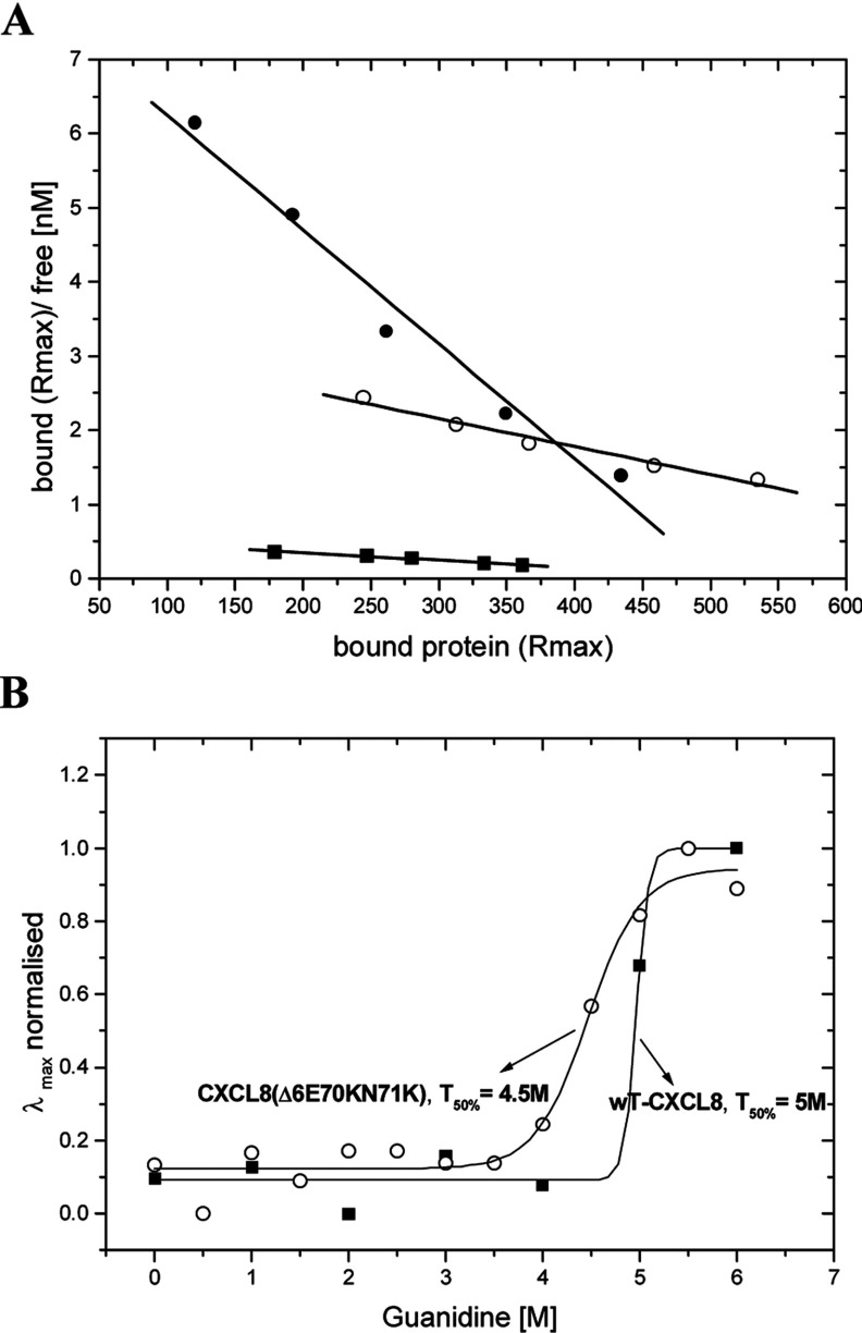
Figure 3. GAG binding and unfolding behaviour of diverse chemokine proteins.
(A) Scatchard plots of HS binding affinities obtained by SPR. wtCXCL8 (■ Kd=1 μM), CXCL8(Δ6E70KN71K) (○ Kd=265nM) and CXCL8(Δ6F17KF21KE70KN71K) (● Kd=65 nM). (B) Guanidine-induced unfolding curves of wtCXCL8 and CXCL8(Δ6E70KN71K). Denaturation was monitored by fluorescence wavelength shift and characterized by the transition midpoint of unfolding T50% [x0 in Eqn. (2)] and by the relative cooperativity [dx in Eqn. (2)].

