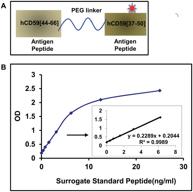
Figure 3. Synthetic Surrogate GCD59 for assay standardization.
A. Diagram of synthetic surrogate standard in which both antigen peptides, hCD59[44-66] and (K41(Nε-glucitol)hCD59[37-50] (hCD59[37-50]), respectively used to raise the capture mouse monoclonal antibody MABTotCD59 and the detection MABGlyCD59 antibodies, were coupled by a PEG linker.
 Represents the pre-formed Nε-glucitollysine residue used to synthesize the (K41(Nε-glucitol)hCD59[37-50] peptide for the position equivalent to K41 in the native protein. The detailed structure of GCD59 synthetic surrogate is shown in Supplemental Figure 3.
B. Characteristic surrogate GCD59 standard peptide dose-response curve in the here described sandwich ELISA that utilizes MABTotCD59 as capture and MABGlyCD59 as detection antibodies.

