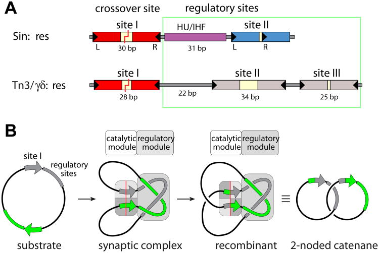
Figure 1. Recombination sites and synaptosome cartoons.
a) Comparison of the full recombination sites for Sin (top) and γδ/Tn3 resolvases (bottom). The Sin site binds two Sin dimers (at sites I and II) and a DNA bending protein (HU or IHF), while the γδ/Tn3 site binds three recombinase dimers. DNA breakage and rejoining occurs Site I. For Sin, the individual subsites are colored to match the proteins bound to them in figure 3.
b) Cartoon of resolution. Synaptosome formation brings two site Is together and traps 3 interdomainal supercoils. A 180° rotation within the site I-bound tetramer creates recombinant products, which are catenated daughter circles. Adapted from [16].

