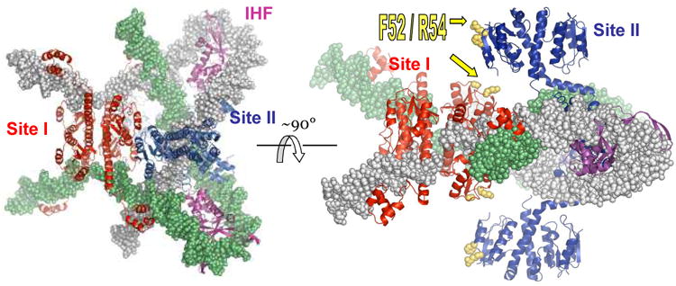
Figure 3.

Model of the Sin synaptosome. Four structures were docked together to create this model: the activated γδ resolvase tetramer - site I complex (red proteins), the IHF-DNA complex (magenta proteins), the Sin – site II complex (blue proteins), and one turn of model-built B-form DNA to continue the path of the DNA past the end of site II [16]. One DNA partner is in gray, the other green. Note that the protein colors correspond to those of their sites in figure 1a.
