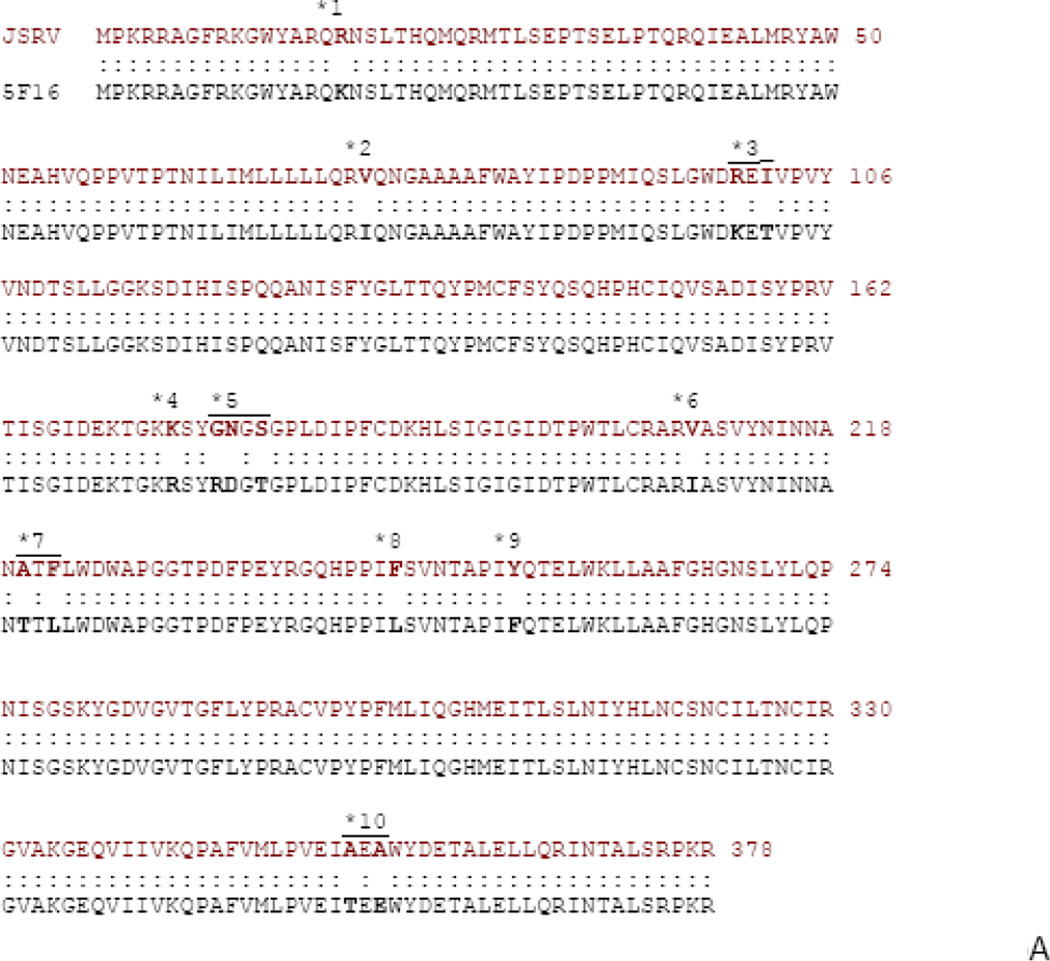
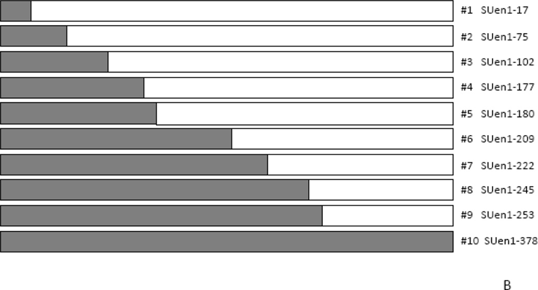
Figure 6. Envelope SU chimeras.
A) Sequence comparison of the Env SU sequences for JSRV and enJS5F16 are shown. Residues where there are differences are shown in bold. The differences are grouped (1 – 10), also shown in the figure. B) The organization of SU chimeras is shown. Site directed mutagenesis was used to sequentially mutate the differing residues starting from the N-terminus, based on ΔGP (JSRV Envex) to give the chimeras indicated.


