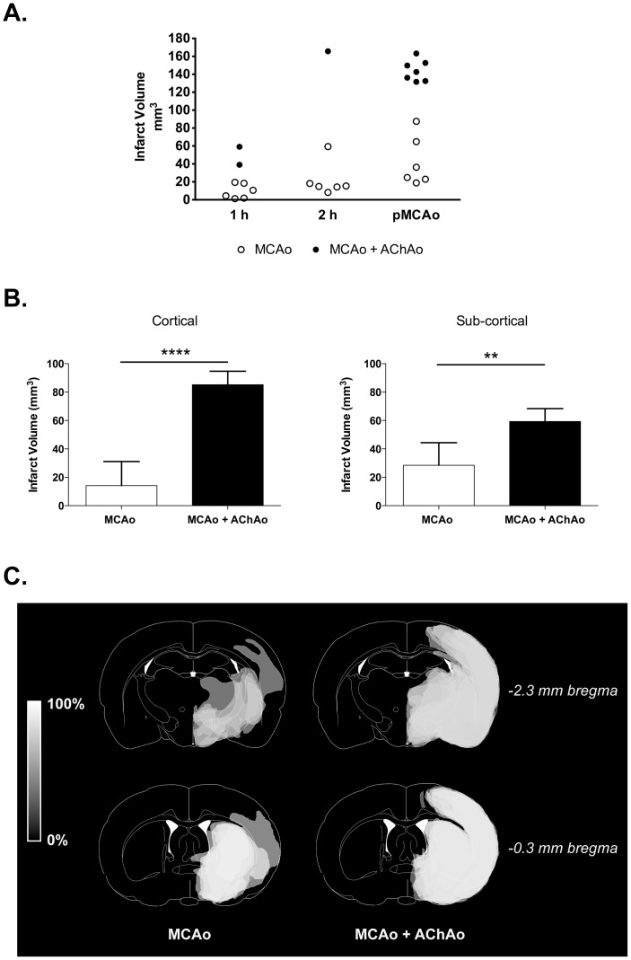
Figure 3. Effect of concurrent anterior choroidal artery occlusion on infarct volume after temporary or permanent MCA occlusion.
Outbred Wistar rats underwent 1 h (n = 8), 2 h (n = 7) or permanent MCAo (n = 13) of the right middle cerebral artery (MCAo) using a silicone-tipped intraluminal filament. A. Infarct volume was quantified with histology at 24 h. Presence of anterior choroidal artery occlusion (AChAo) was determined by cerebral blood volume (CBV) map analysis. B. Cortical and sub-cortical infarct volume distribution in pMCAo animals (mean + sd). C. The traced infarct (seen in grey) from animals in the pMCAo group were overlaid so that lighter regions represent areas more commonly infarcted.

