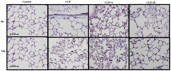
Figure 2. ICAM-1 staining in lung.
CLP was induced in mice (n = 8 in each group), there were three groups: 1- only CLP; 2- 30 min later treated with hypertonic saline 7.5% (CLP+H) or 3- treated with normal saline solution 0.9% (CLP+S). Forth group (C) was used to indicate basal levels. Samples from lung (a) were harvested after 6, 12 and 24 h. There was a positive staining for ICAM-1 in CLP animals, showing the presence of inflammation activating molecules for neutrophil recruitment. There was no difference in the intensity of ICAM-1 staining between CLP and CLP treated with normal saline. Hypertonic solution reduced ICAM-1 activation in CLP mice.

