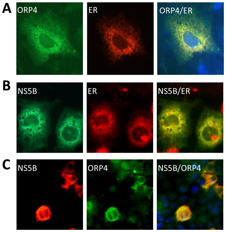
Figure 3. Expression and subcellular localization of ORP4.
Huh7.5 cells were transfected with pCMV-flag-ORP4 and pDsRed2-ER (A), pCMV-myc-NS5B and pDsRed2-ER (B), or pCMV-flag-ORP4 and pCMV-myc-NS5B (C) expressing an ER-targeted red fluorescence protein (RFP). Forty-eight hours post-transfection, cells were washed, fixed with 4% PFA, permeabilized and stained with anti-flag antibody for ORP4 or anti-myc for NS5B, followed by FITC-conjugated IgG for ORP4 (A & C), Alexa-fluor 488 IgG for NS5B (B) or PE-conjugated IgG for NS5B (C). Appropriate isotype-matched IgG antibodies were included to ensure the antibody specificity. Nuclei of the cells were stained with DAPI. Representative micrographs of stained cells were taken using a Zeiss M200 fluorescence microscope. The data were representative of three independent experiments.

