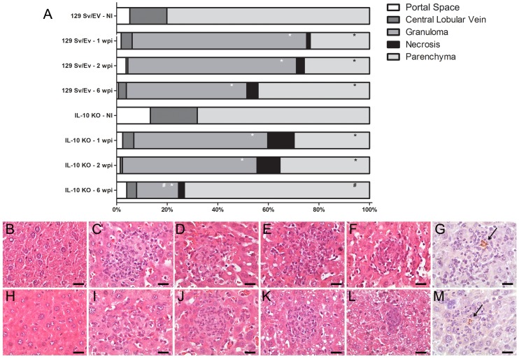
Figure 5. Morphometric analysis, histopathology and immunohistochemistry of hepatic tissue of B. abortus infected IL-10 KO mice.
(A) Columns indicate volumetric proportions of tissue components. The number of portal space, central lobular vein, granuloma, necrosis and parenchyma were evaluated and transformed in percentage at 1, 2 or 6 weeks postinfection. Statistically significant differences relative to non-infected group (NI) are represented by an asterisk (p<0.05). Differences relative to granuloma number from IL-10 KO mice compared to wild-type mice at six-week postinfection are indicated by #. Similar results were obtained in two-independent experiments. (B-F) Representative of hematoxylin- and-eosin-stained sections of hepatic tissue from wild-type mice uninfected (B) or infected at one- (C), two- (D), three- (E) or six-weeks (F). (H-L) Representative of hematoxylin- and eosin-stained sections of hepatic tissue from IL-10 KO mice uninfected (H) or infected at one- (I), two- (J), three- (K) or six-weeks (L). Immunohistochemistry sections of hepatic tissue from wild-type (G) and IL-10 KO (M) mice containing the B. abortus inside the granuloma. The arrows indicate the B. abortus within the granuloma. Scale bars: 20 µm.

