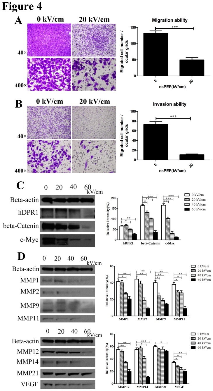
Figure 4. NsPEF inactivates cancer cells metastasis and invasion by inhibiting Wnt/β-Catenin signaling pathway to down-regulate expressions of VEGF and MMPs family proteins in vitro.
(A) Migration ability of cancer cells exposed to nsPEF was tested by trans-well assay. The migrated cells exposed to nsPEF were stained purple by 0.1% crystal violet solution, observed under light microscope for 40 or 400 magnifications, and counted for statistical analysis. Original magnification, 40× & 400×. ***p<0.001. (B) Invasion ability of cancer cells exposed to nsPEF was detected using matrigel invasion assay. After nsPEF treatment, the cells that possessed invasion ability penetrated through the matrigel, were stained purple by 0.1% crystal violet solution, and were counted for statistical analysis. Original magnification, 40× & 400×. ***p<0.001. (C) Protein expressions of Wnt/β-Catenin signaling pathway including hDPR1, β-Catenin and c-Myc in cancer cells after exposure to nsPEF with different intensities were detected by Western-blot assay. (D) Protein expressions of MMPs family and VEGF in cancer cells after exposure to nsPEF with different intensities were detected by Western-blot assay. Relative intensity of each protein was analyzed by Photoshop CS4 software. *p<0.05, **p<0.01, ***p<0.001.

