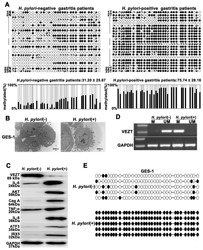
Figure 2. Helicobacter pylori infection promotes the methylation and silencing of VEZT.
(A) H. pylori-positive gastritis patients showed higher methylation levels in the VEZT promoter region when compared with H. pylori-negative gastritis patients. Each row of circles represents an integrated methylation ratio from three clones, and each circle represents a single CpG site. Open circle represents unmethylated cytosine, whereas filled circles or partially filled circles represent the methylated ratio of CpG sites. (B) After 24-h infection with H. pylori, the attachment of H. pylori was observed by transmission electron microscopy on the surface of GES-1 cells in the experimental cells relative to the control cells. (C) VEZT expression level in GES-1 cells was reduced after a 24-h H. pylori infection relative to negative control cells; however, H. pylori infection induced the IL-6, AKT, TNF-α, IL-8, ATF3 and IRX5 expression by western blot analysis. (D) The methylation of the VEZT promoter was detected by MSP after a 24-h H. pylori infection in GES-1 cells (marked as M), whereas methylation of the VEZT promoter in GES-1 cells that were not infected with H. pylori was not observed (marked as U). (E) Schematic summary of 34 CpG sites in the promoter region of the VEZT gene from -171 to -428 by BSP analysis. GES-1 cells showed a higher level of methylation after H. pylori infection relative to the control samples. The bar represents 5000 nm.

