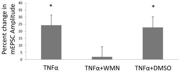
Figure 3. Blockade of the TNFα-induced increase in mEPSC-amplitude by wortmannin (a PI-3-K inhibitor).

Shown are percent changes in amplitude of mEPSCs after TNFα (60-ng/ml) (TNFα), TNFα (60 ng/ml) with 1 μM wortmannin (WMN) incubation for 30-minutes (TNFα + WMN), and TNFα (60 ng/ml) with 0.1% DMSO incubation for 30 minutes (TNFα + DMSO; a control). For all treatments, F(2,16) = 5.07, p < 0.05; n = 6–7. Post hoc LSD test shows that the percent change of the mEPSC-amplitude by TNFα. TNFα + WMN-exposure is significantly less than the change in amplitude after TNFα alone (p<0.05) and the TNFα with DMSO (p<0.05).
