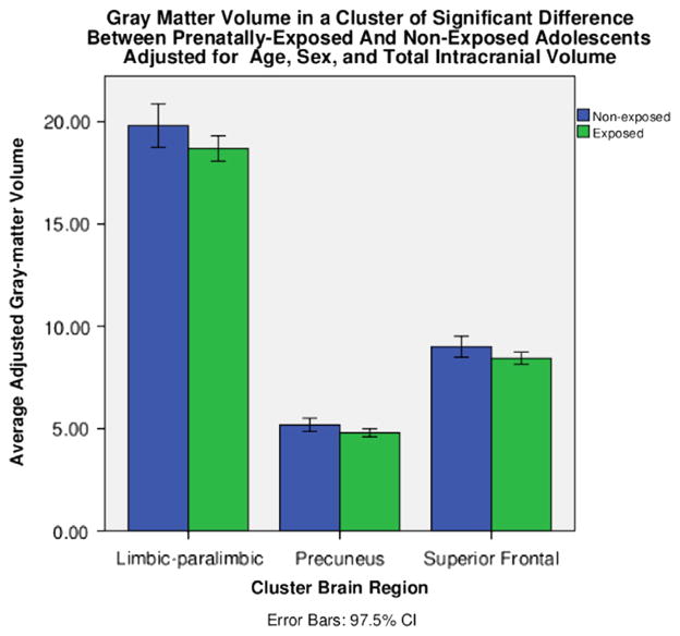
Figure 2.
Average gray matter volume in milliliters by group for each of the three regions of significantly lower gray matter volume in prenatally cocaine-exposed than noncocaine-exposed adolescents (ps < .025 family-wise error corrected). From left to right: limbic and paralimbic cortex; parietal cortex, including the superior parietal lobe and precuneus region; and superior frontal cortex. CI, confidence interval.

