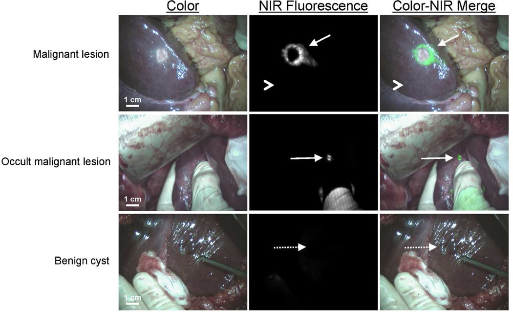
Figure 1. NIR fluorescence imaging of colorectal liver metastases.
A colorectal liver metastasis (arrow) is clearly identified by a rim around the tumor in vivo (top row), 24 h after injection of 10-mg ICG. Normal liver tissue (arrowhead) shows minimal background uptake of ICG. In 5 patients, small, superficial, otherwise occult metastases (middle row, arrow) were identified by NIR fluorescence imaging. Benign lesions (bottom row, dashed arrow) could be differentiated from malignant lesions by a lack of a fluorescent rim around the lesion.

