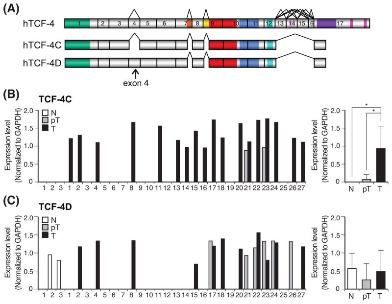
Fig. 1. Expression profiles of TCF-4C and D mRNA in human HCC samples.
(A) A schematic representation of human TCF7L2 gene compromising 17 exons based on the reported cDNA sequences (36) and structural organization of human TCF-4C and TCF-4D isoforms. The alternatively spliced sites, shown with a tee-pee shape, are exon 4, 7L (LVPQ), 9L (SxxSS), 13, 14, 15, 16, and 17L. (B, C) Expression level of TCF-4C (B) and TCF-4D (C) measured by semi-quantitative RT-PCR in human HCC tumors (T), adjacent peritumor tissue (pT) and normal liver (N). Left panel shows expression level in each sample, and right panel depicts as mean ± SD. The expression level of TCF-4C and TCF-4D was evaluated by semi-quantitative RT-PCR and the values were normalized by GAPDH. *, P < 0.05.

