4.
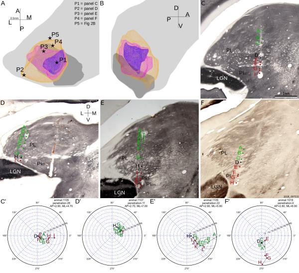
The representation of the central-peripheral axis of the visual field. A-B: Horizontal (panel A) and parasagittal (panel B) views of the representations of visual field areas within 5 degrees (blue), 10 degrees (pink) and 15 degrees (yellow) of the area centralis. Same conventions as Figure 3. C-F: Reconstructions overlaid on coronal CO sections, of example penetrations whose locations are shown in panel A, with same conventions as in Figure 2. C'-F': perimeter charts of penetrations shown in panels C-F. PIcm, medial part of central inferior pulvinar; Picl, lateral part of central inferior pulvinar.

