
Fig.1. GCV induces selective cell killing in MSC-TK.
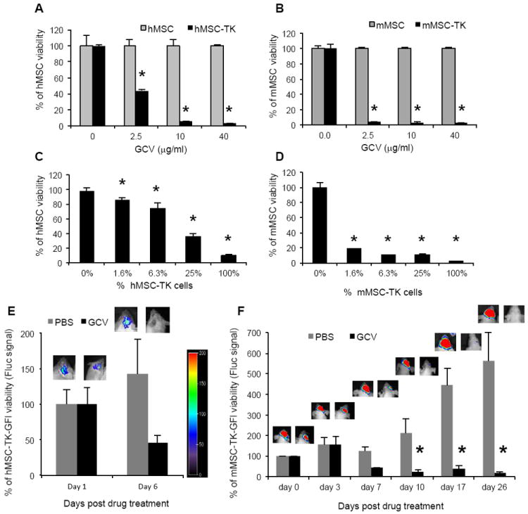
(A-B) hMSCs (A) or mMSC (B) were transduced with LV-TK or RV-TK respectively and 48hrs later were plated and incubated with serial dilutions of GCV (0-40 μg/mL). Plots show MSC viability at 72 hrs post GCV treatment. Bars, +SD. (C-D) Different proportions of hMSC-TK (C) or mMSC-TK (D) were plated with non-transduced hMSC or mMSC respectively and incubated with GCV (10μg/mL). Plots show MSC viability 72hrs post GCV treatment. Bars, + SD. All experiments were performed in triplicates and repeated twice. (E-F) hMSC-TK-GFl (E) or mMSC-TK-GFl (F) were implanted intracranially in adult SCID or nude mice respectively and treated with GCV (50mg/kg). Plots show percentage of MSC viability measured by Fluc intensities of control PBS and GCV group. Bars, +SE. One representative image of a mouse from each group and from each time point is shown. In all panels, *, p < 0.05 versus controls. Abbreviations: hMSC, human mesenchymal stem cells; mMSC, mouse mesenchymal stem cells; GCV, ganciclovir; GFl, green fluorescent protein and firefly luciferase fusion; TK, Thymidine Kinase; PBS, phosphate buffered saline.