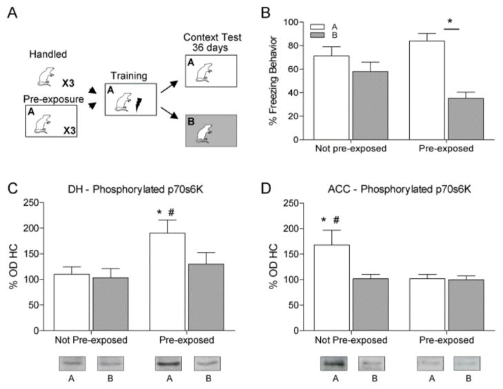
Figure 2.
(A) Rats were handled or pre-exposed to the training chamber for 3 days, trained the next day, and tested 36 days later in either context A (white) or B (gray). (B) Only rats given pre-exposure significantly discriminated between contexts (* p < 0.05). (C) Western blot analysis showed a significant p70s6K increase in the DH when animals were pre-exposed and placed in Context A for testing compared to HC, pre-exposed B, or the not pre-exposed Context A group. (D) In contrast there was a significant increase in phosphorylated p70s6K in the ACC in rats not pre-exposed and tested in A compared to HC, animals not pre-exposed and tested in B or pre-exposed and tested in Context A.

