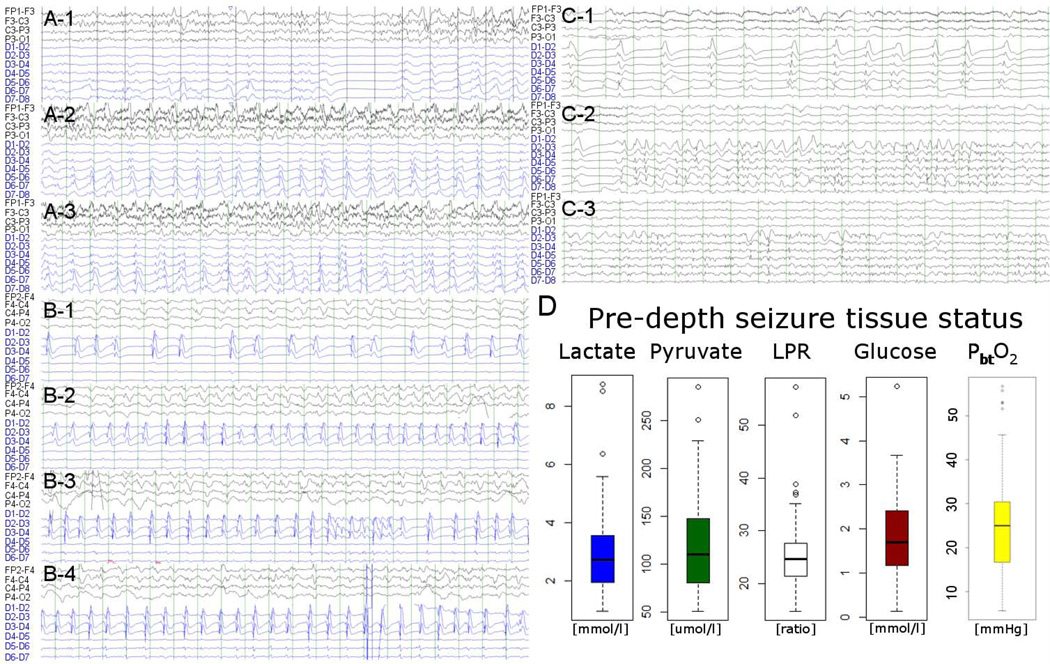
Figure 1.
Three illustrative intracortical seizure cases (A through C) are displayed (ipsilateral scalp EEG each in the top 4 and minidepth EEG in the bottom 6 to 7 channels; all bipolar montage). Case A intracortical seizure with surface seizure: 54 year old woman with poor grade SAH (Hunt Hess 5, APACHE-2 27), that underwent clipping of an anterior communicating artery aneurysm. Case B intracortical seizure with ictal-interictal continuum surface recording: 47 year old man with poor grade SAH (Hunt Hess 5, APACHE-2 14), that underwent clipping of a right anterior cerebral artery aneurysm. Case C intracortical seizure with non-ictal patterns on surface recordings: 57 year old woman with poor grade SAH (Hunt Hess 4, APACHE-2 17), that underwent clipping of an anterior communicating artery aneurysm. Right lower panel shows baseline tissue status based on microdialysis and partial brain tissue oxygenation averaged for all patients over 60 minutes prior to intracortical seizure onset indicating an overall non-ischemic state (median glucose 1.7mmol/L [IQR 1.2–2.4], pyruvate 110umol/L [IQR 80–148], lactate 2.8mmol/L [IQR 1.9–3.6], LPR 25 [IQR 21–28], PbtO2 24.4mmHg [IQR 4.7–63.0]). Only two events were preceded by an LPR >40 and 9 had a cerebral glucose of 0.7 of below, no event was preceded by metabolic crisis (LPR > 40 and brain glucose < 0.7mmol/L).

