
Figure 6. Efficacy of AAV2 in vivo across different brain areas.
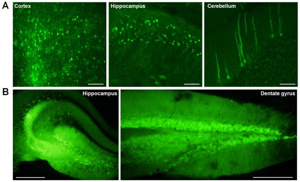
To test efficacy of AAV2 in vivo, P0 or stereotaxic injection of mice was performed and expression of GFP was qualitatively evaluated four weeks post-injection. (A) Representative images of P0 injection of AAV2. Extensive spread of GFP fluorescence was observed in multiple brain regions, including (left to right) cortex, hippocampus, and cerebellum. Scale bars = 100 μM. (B) Representative images of stereotaxic injection of AAV2. GFP fluorescence was observed in all hippocampal regions. Image on right is more posterior. Scale bars = 500 μM. Data representative of three mice.