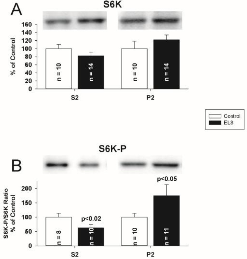
Figure 10. ELS does not mediate alterations in localization of S6K, but does mediate changes in S6K–P at P60.
A, S6K was not significantly removed from the cytosolic compartment, though a trend similar to PP2A and FRMP was suggested (S2 fraction, ELS: 82.01 ± 9.45%, n = 14; control: 100 ± 10.50%, n = 10, P = NS, Student’s t-test). S6K P2 (synaptosomal) fraction was not changed (ELS: 121.90 ± 12.18%, n = 14, control: 100 ± 18.58%, n = 10, P = NS, Student’s t-tests). B, Phosphorylated S6K (Thr 389) was significantly reduced in the cytosolic compartment (S2 fraction, ELS: 63.13 ± 10.35%, n = 10; control: 100 ± 13.16%, n = 8, P < 0.02, Student’s t-test). Phosphorylated S6K (Thr 389) was significantly increased in the P2 (synaptosomal) fraction (ELS: 175.00 ± 38.58%, n = 11, control: 100 ± 13.04%, n = 10, P < 0.05, Student’s t-tests). Semi-quantitative western-blot technique(Cornejo et al., 2007) was used to determine immunoreactivity/mg loaded protein, which were normalized to controls (see Methods). Typical blots with equivalent protein loading shown. Solid bars ELS, open bars control.

