
Figure 4. Abi3bp regulates cyclin-d1 expression and ERK phosphorylation.
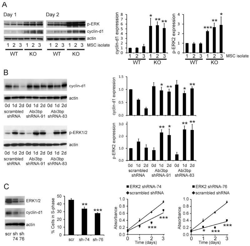
(A) Extracts (7.5μg) from wild-type and Abi3bp knockout MSC isolates, one and two days post seeding, were immunoblotted for p-ERK1/2 and cyclin-d1. Actin was used as a loading control. Intensities were normalized to actin; normalized intensity of wild-type isolation 1 was taken to be 1. (B) Extracts (7.5μg) of MSC-GFP-Akt1 cells expressing either control scrambled or Abi3bp shRNA at zero, one, and two-days post-seeding were probed for cyclin-d1 or phospho-ERK1/2. Actin was used as a loading control. Intensities were normalized to the loading control; normalized intensity of scrambled control cells at day 0 was taken to be 1. N=3 (cyclin-d1), N=4 (p-ERK). Comparisons between groups at the same time point *p≤0.05, ** p≤0.01. (C) Stable knockdown of ERK2 by shRNA. Far left panel: immunoblotting ERK n=4. Left panel: Cells were incubated with BrdU for 6 hours one day post seeding. Percentage of cells in S-phase determined by flow cytometry. N=4. Comparisons between knockdown and control cells ** p≤0.01, *** p≤0.001. Right panels: MTS assay growth curves. N=5. Comparisons between groups at the same time point *p≤0.05, *** p≤0.001.