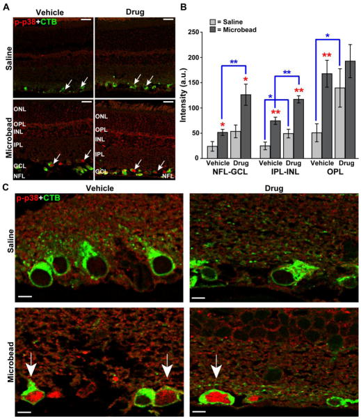
Figure 3. Ro3206145 does not prevent p38 MAPK activation, as expected.
A) Immuno-labeling for phosphorylated p38 MAPK (p-p38) in retinal sections from either saline (top row) or microbead (bottom row) eye from study cohorts. Six weeks of microbead-induced elevated IOP increased signal, especially near RGCs labeled by uptake of cholera toxin B (CTB, arrows), which was injected into the eye prior to sacrifice; this is not affected by Ro3206145 treatment. Abbreviations: ONL (outer nuclear layer), OPL (outer plexiform layer), INL (inner nuclear layer), IPL (inner plexiform layer), GCL (ganglion cell layer), and NFL (nerve fiber layer). B) Quantification of phosphorylated p38 MAPK signal across retinal layers in arbitrary units (a.u.; n≥7 sections per cohort). Elevated IOP increased signal most significantly near RGCs and their axons (NFL-GCL) and in the OPL. Drug treatment tended to exacerbate this increase. * p ≤ 0.05 or ** p ≤ 0.01 for microbead vs. saline retina (red) and drug vs. vehicle cohort (blue brackets). C) High magnification micrographs show increased phosphorylated p38 MAPK with elevated IOP in the nucleus of CTB-labeled RGCs (arrows) for both vehicle and drug cohorts. Scale = 20 μm (A), 5 μm (C).

