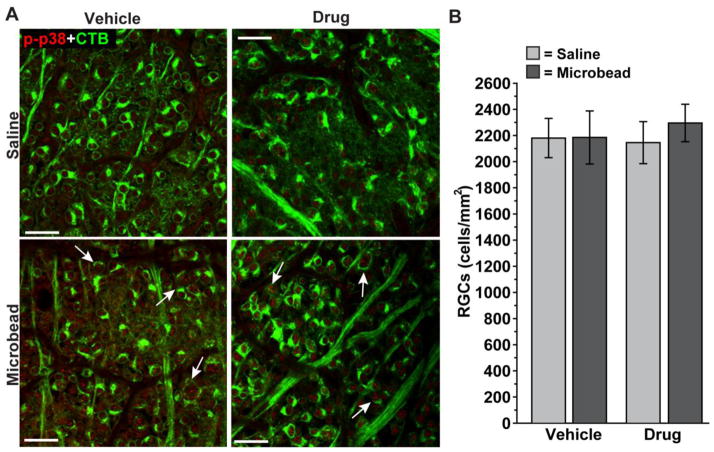
Figure 6. Rescue of axon transport by p38 MAPK inhibition is not due to changes in CTB uptake.
A) Confocal micrographs through the nerve fiber and ganglion cell layers of whole-mounted retina from vehicle and drug cohorts showing CTB-labeled RGCs and their axons. As in Figure 3, retina from microbead-injected eyes demonstrate increased phosphorylated p38 MAPK (p-p38) in both cohorts; arrows indicate particular examples of CTB-labeled RGCs with p-p38 localizing in the nucleus. Scale = 20 μm. B) Quantification indicates similar numbers of CTB-labeled RGCs between cohorts, for both saline and microbead retina. Thus, differences in anterograde transport to the SC as shown in Figure 4 are not due to differences in CTB uptake by RGCs. n ≥ 3 sets of retina per cohort.

