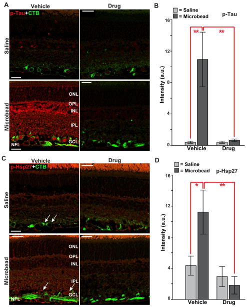
Figure 7. Ro3206145 reduces downstream target activation.
A) Immuno-labeling for phosphorylated Tau (p-Tau) in retinal sections from either saline (top row) or microbead (bottom row) eye from study cohorts. Signal is increased throughout the retina in the vehicle cohort with microbead-induced elevated IOP; drug treatment prevents this. CTB label in RGCs included for reference. B) In the vehicle group, intensity of phosphorylated Tau signal in microbead retina was increased 10-fold (10.9 ± 3.5 vs. 0.4 ± 0.1 for saline retinas; mean ± sd). C) Immuno-labeling for phosphorylated Hsp27 (p-Hsp27) also shows increased levels in retinal sections with elevated IOP in the vehicle cohort, especially near RGCs (arrows). Increase was prevented in the drug cohort. D) For retinas from the vehicle group, elevated IOP increased phosphorylated Hsp27 three-fold (11.2 ± 2.8 vs. 4.3 ± 1.2 for saline eye). n ≥ 7 images per cohort; * p ≤ 0.05, ** p ≤ 0.01. Abbreviations as in Figure 3. Scale = 15 μm.

