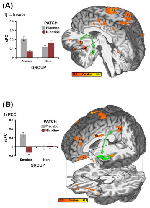
Figure 4.
rsFC strength in amygdala- and insula-centric circuits was decreased by nicotine in abstinent smokers but not in nonsmokers. (A) A GROUP x PATCH interaction analysis identified brain regions whose rsFC with a left amygdala seed (S, green: same as in Fig. 2A) showed differential responses to nicotine challenge in smokers versus nonsmokers. Qualitatively, nicotine decreased rsFC between the amygdala and insula (1) in the smoker but not the nonsmoker group. Note: This whole-brain between-group strategy independently identified similar regions as those detected in the within-smoker analysis (c.f., Fig. 2A). Similar interaction patterns were observed in the precentral gyrus (2), parietal regions (3), and posterior insula (4); see also Fig. S6 and Table S4. (B) A GROUP x PATCH interaction analysis identified brain regions whose rsFC with a left insula seed (S, green: same as in Fig. 2B) showed differential responses to nicotine challenge in smokers versus nonsmokers. Nicotine decreased rsFC between the insula and PCC/precuneus (1) in the smoker but not the nonsmoker group. Similar interaction patterns were observed in the vmPFC (2), dmPFC (3), and mid-cingulate cortex (4); see also Fig. S7 and Table S5.

