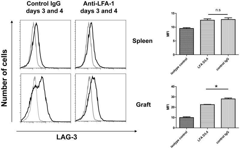
Figure 7. Expression of LAG-3 on CD8 T cells in spleens and cardiac allografts from recipients treated with anti-LFA-1 mAb.
Groups of C57BL/6 mice were treated with 200 μg control rat IgG or anti-LFA-1 mAb on days 3 and 4. The mice received complete MHC mismatched A/J cardiac allografts on day 0. Recipient spleens (top rows) and allografts (bottom rows) were harvested on 7 post-transplant, digested to prepare single cells suspensions, and aliquots stained with anti-LAG-3 (solid line) or control IgG (shaded line) and anti-CD8 mAb and analyzed by flow cytometry to assess LAG-3 expression by CD8 T cells in the recipient spleen and by the graft infiltrating CD8 T cells. Representative histograms of LAG-3 expression by gated CD8 T cells in grafts from each group is shown. The mean change in Mean Fluorescence Intensity (MFI) of LAG-3 staining for the gated CD8 T cells in 4 grafts from each group SEM is shown. *p ≤ 0.03; n.s. not significantly different.

