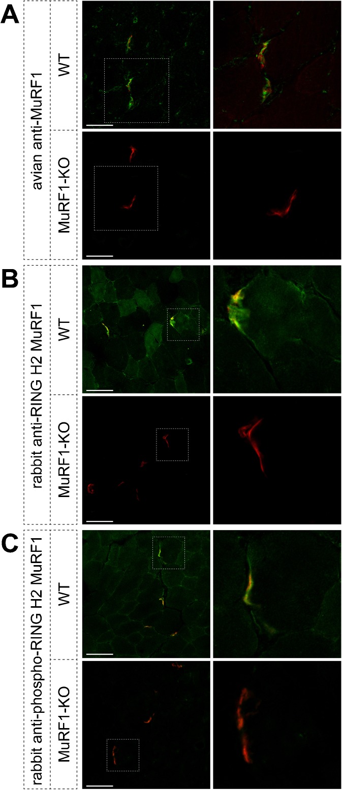
Fig. 1.
Endogenous MuRF1 is highly enriched in close proximity to the NMJ. EDL muscles of WT and MuRF1-KO animals were sectioned transversally and co-stained with the AChR marker, BGT-AF555, and with three different anti-MuRF1 antibodies, i.e., IgY-type from chicken against the MuRF1 coiled-coil domain (a); IgG-type rabbit polyclonal antibodies directed to the MuRF1 RING H2 domain (b, c). Fluorescence distribution was then determined using confocal microscopy. Pictures show single optical sections of fluorescence signals of BGT-AF555 (red) and anti-MuRF1 (green) of WT and MuRF1-KO muscles, as indicated. Right panels depict high power views of the boxed areas of the images on the left. Scale bars represent 50 μm

