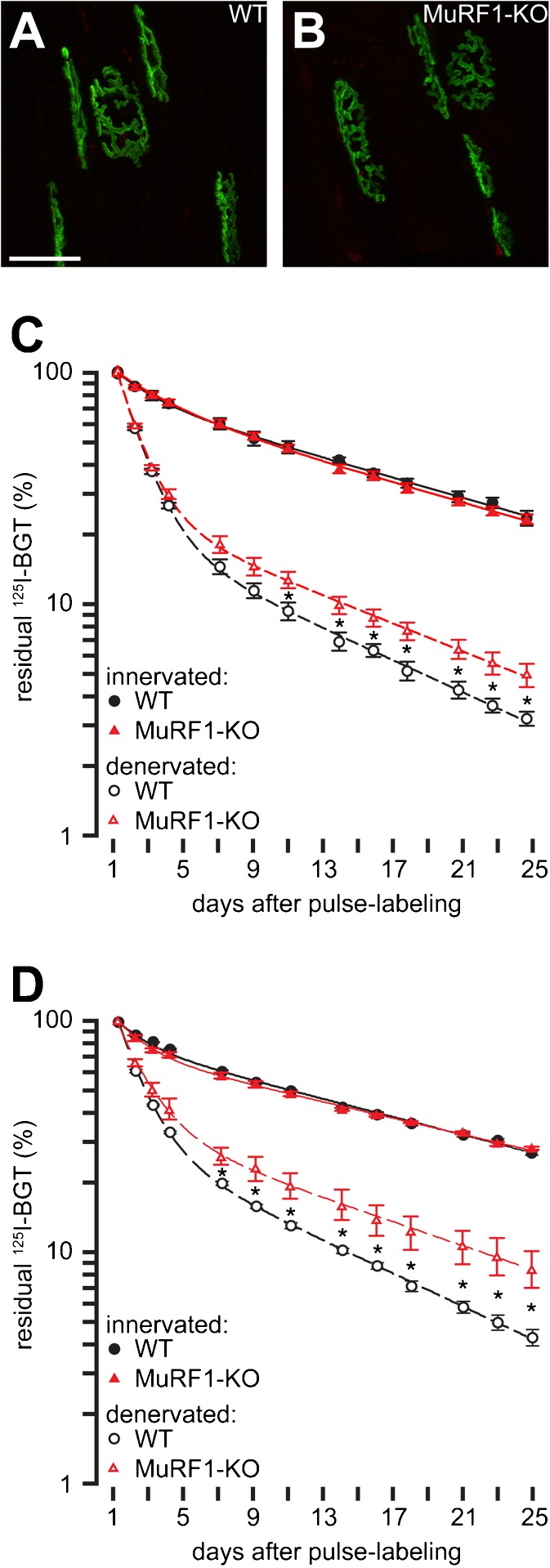
Fig. 3.
Metabolic destabilization of AChR upon denervation is partially rescued in MuRF-1KO animals. a Tibialis anterior muscles of wild-type (WT) and b MuRF1-KO animals were injected with infrared fluorescent BGT-AF647 to label AChRs present at that time point ('old receptors'). Ten days later, red fluorescent BGT-AF555 was injected to mark 'new receptors' and then muscles were imaged with confocal microscopy. Panels show maximum-z projections of 'old receptors' and 'new receptors' in green and red, respectively. Note, that all NMJs displayed hardly any 'new receptor' signals. Left legs of WT and MuRF1-KO animals were denervated, right legs served as innervated controls (c) or were left untreated and innervated controls were from separate animals (d). Five days later, AChRs in tibialis anterior muscles of all legs were pulse-labeled with 125I-BGT. Then, 125I emission was monitored at indicated intervals during the next 4 weeks and normalized to the values measured 24 h after pulse labeling. Symbols show measured values (mean ± SEM, c: n = 3 for WT and n = 4 for MuRF1-KO, d: n = 4 for both denervated WT and denervated MuRF1-KO muscles and n = 5 for both innervated WT and innervated MuRF1-KO muscles). Lines depict two-term exponential fits. *p < 0.05

