Fig. 5.

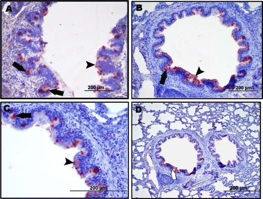
a–d Positivity for M. ovipneumoniae antigens on the surface (arrow head) and cytoplasm (arrows) of bronchial and bronchiolar epithelial cell. Immunoperoxidase staining, ABC

a–d Positivity for M. ovipneumoniae antigens on the surface (arrow head) and cytoplasm (arrows) of bronchial and bronchiolar epithelial cell. Immunoperoxidase staining, ABC