
Figure 4.
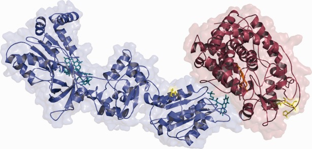
Structure of the CYPOR-2D6 complex after 10 ns. CYPOR is shown in blue and 2D6 in red. The positions of the membrane anchors and the two leucines which should be embedded in the membrane are shown in yellow. [Color figure can be viewed in the online issue, which is available at http://wileyonlinelibrary.com.]